
MBBS (AIIMS), MS (Surgery, AIIMS), MNAMS, FACS (USA), FICS (USA), FUICC
Mon, 21 Sep 2026
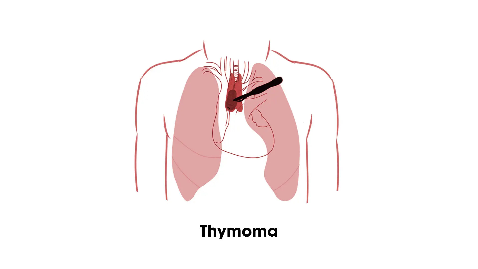
A diagnosis of thymoma can feel overwhelming, especially since most people have never heard of the thymus gland before receiving one. Common questions follow quickly — is it cancerous, does it spread, is surgery necessary, and where should treatment be taken.
Dr. Arvind Kumar, Chairman of the Institute of Chest Surgery at Medanta, treats thymoma and other mediastinal tumors using robotic and minimally invasive surgical techniques.
India has grown into a well-established destination for thoracic oncology care, supported by experienced medical professionals, modern infrastructure and treatment costs that remain considerably lower than in the US, UK or Australia.
Thymomas are often called “silent” tumors — they develop in the thymus gland, a small organ in the upper chest behind the breastbone, and may not cause symptoms in their early stages. For the most part, they are discovered incidentally during a chest X-ray or CT scan done for unrelated reasons. When symptoms do occur, they are typically the result of the tumor pressing on nearby structures. A more detailed breakdown is available in our guide to thymus cancer symptoms and causes.
Common symptoms of thymoma include:
Approximately 30% to 50% of thymoma patients also have Myasthenia Gravis (MG), an autoimmune disorder in which the body’s immune system mistakenly attacks the junction between nerves and muscles, causing muscle weakness. In most cases, a thymectomy — removal of the thymus — meaningfully improves MG symptoms and helps restore quality of life.
The first step in thymoma treatment is a thorough diagnosis. The following diagnostic tools are commonly used for thymoma detection, explained further in our guide to thymus cancer diagnosis:
Staging is the next crucial step in planning treatment.
The treatment pathway for thymoma depends heavily on its stage. In India, surgeons generally follow the Masaoka-Koga staging system, which categorizes the tumor based on its level of invasion.
| Stage | Description | 5-Year Survival Rate (Avg.) |
|---|---|---|
| Stage I | Completely encapsulated; no invasion of the capsule. | ~95% |
| Stage II | Microscopic invasion into the surrounding fat or pleura. | ~86% |
| Stage III | Macroscopic invasion into neighboring organs (lungs, heart, great vessels). | ~41%–70% |
| Stage IV | Spread to the pleura, pericardium, or distant organs. | Varies by case |
As with most tumors, early detection matters for thymoma. Diagnosis at Stage I or II allows for complete surgical resection, which meaningfully increases the chances of a definitive cure.
Thymoma is not treated like a routine chest condition, primarily because of its location around vital structures, which makes the treatment approach delicate. The thymus lies close to:
This means that even a small tumor can create complications due to its position near the mediastinum. This is one of the reasons why consulting an experienced thymoma specialist is important.
Surgery remains the primary treatment for thymoma. Initially, a median sternotomy was the most commonly used surgical route. Medical advancements have since introduced more refined and precise surgical options.
India has seen a significant shift toward robotic thymectomy. Using the Da Vinci Robotic System, the entire procedure can be performed through three or four small incisions (8–10mm each). Reasons for its growing use include:
VATS is another minimally invasive, keyhole surgical technique. While VATS is highly effective, robotic surgery is often preferred for thymoma because of the robot’s greater manoeuvrability in the tight space of the anterior mediastinum.
For advanced thymomas (Stage III and IV), surgery alone may not be enough, so a multidisciplinary approach is used:
The treatment protocol is personalized based on a comprehensive evaluation and diagnosis.
Thymoma can often be treated successfully, particularly when diagnosed early. Complete surgical removal of the tumor is the most important factor influencing long-term outcomes. Most patients diagnosed at Stage I and II respond well to surgery alone or surgery combined with radiation therapy; some advanced cases also respond well to multimodal treatment involving surgery, chemotherapy, and radiation. Even after successful tumor removal, long-term monitoring is needed to watch for possible recurrence.
Early diagnosis and treatment of thymoma are important. Untreated thymoma may:
Early intervention allows for safer surgery and better outcomes. If you are experiencing unexplained chest symptoms or persistent muscle weakness, consult a thymoma specialist promptly for evaluation.
Most patients gradually return to normal life after successful treatment, though long-term monitoring remains important. Follow-up care typically includes:
Beyond physical health, a cancer diagnosis can affect emotional well-being too. Supportive counselling, family involvement, and rehabilitation are part of ongoing care as needed.
Advancements in robotic surgery and thoracic oncology have made the path to recovery clearer for thymoma patients. Early intervention supports better outcomes and a faster return to normal life. If you have been diagnosed with a mediastinal mass or thymoma, Dr. Arvind Kumar and his team at the Institute of Chest Surgery provide personalized, patient-centric care — from diagnosis through surgery and long-term follow-up.
For more details about thymoma treatment and to schedule a consultation, please get in touch with our team directly.

Dr. Arvind Kumar is a highly accomplished Chest surgeon with experience of over 20,000 major chest surgeries, including open surgery, VATS(Video assisted Thoracic Surgery), Robotic Chest Surgery, and Lung Transplantation, reflecting decades of clinical excellence and leadership in chest surgery.
Copyright @ (Prof.) Dr. Arvind Kumar. All Rights Reserved / Thoracic Surgical Oncologist
License Number: U.P State Medical Council (India) No. 27637


 Contact
 +91-9773635888