
MBBS (AIIMS), MS (Surgery, AIIMS), MNAMS, FACS (USA), FICS (USA), FUICC
Wed, 05 Jul 2023
.webp)
Despite being very uncommon, chest wall tumours can comprise a variety of benign and malignant tumours that arise inside the chest wall, which is made up of the ribs, muscles, and connective tissues. These tumours can develop from a variety of chest-related tissues, giving rise to a wide variety of manifestations and subsequent problems that can be heightened by a variety of external and internal factors.
Chest Wall tumors usually cause no symptoms and present as a lump growing in the chest wall. Tumors of the chest wall include those that grow on the ribs and sternum. These can be both malignant and benign.
Primary Chest Wall Tumours: These malignancies develop directly inside the chest wall. Malignant (cancerous) or benign (non-cancerous) ones are both possible. Chondrosarcoma, osteosarcoma, and Ewings sarcoma are a few examples.
Secondary Chest Wall Tumours: Also referred to as metastatic tumours, these malignancies spread from other body areas, such as the lungs, breasts, or bones, to the chest wall.
Read More :- Dr. Arvind Kumar
Depending on where they are, how big they are, and whether or not they are benign or malignant, chest wall tumours can present with a wide range of signs and symptoms.
Chest Wall tumors usually cause no symptoms and present as a lump growing in the chest wall. Occasionally it may cause local pain or even fractures of the ribs.
It takes a systematic strategy that combines clinical assessment, imaging, and tissue collection to diagnose chest wall tumours. The diagnosis is confirmed by a carefully performed biopsy.
The goal of surgery for chest wall tumours is to remove the tumour while keeping the patients breathing and chest wall function intact. A multidisciplinary team of surgeons, oncologists, and other professionals frequently performs surgery on chest wall tumours. The objective is to completely remove the tumour while retaining as much of the normal function of the chest wall as is feasible. The precise surgical strategy is determined by the tumours size, location, and malignant or benign nature.
It is usually important for patients to recover and undergo rehabilitation judiciously, and they could need physical therapy to fully restore their strength and flexibility. Each patients personal needs will be taken into account while determining the surgical strategy and after-care schedule.
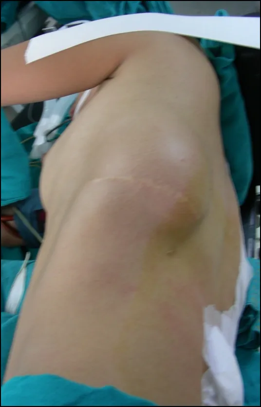
The treatment for chest wall tumors is done by surgery in which complete removal of full thickness of chest wall including all the muscles and ribs with more than 2 cm clear margin around the tumor and then reconstruction of the chest wall by using various imported synthetic materials, including complex plastic surgery operations.

Patients experience | Huge Chest Wall Tumour Surgery

Prof. (Dr). Arvind Kumar honoured with "Fakhr-e-Hind" (Pride of Nation) award in an International event "Jashn-e-Bharat" organised by Indian Heritage and Health Care Centre and Husanara Trust on 27th January 2019 at India International Centre, Delhi.
A lung transplant is a life-saving and complicate...
Lung transplantation is an extremely sensitive a...
Choosing the best team for a lung transplant can...
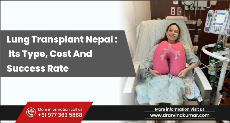
A lung transplant is a surgery which is performe...
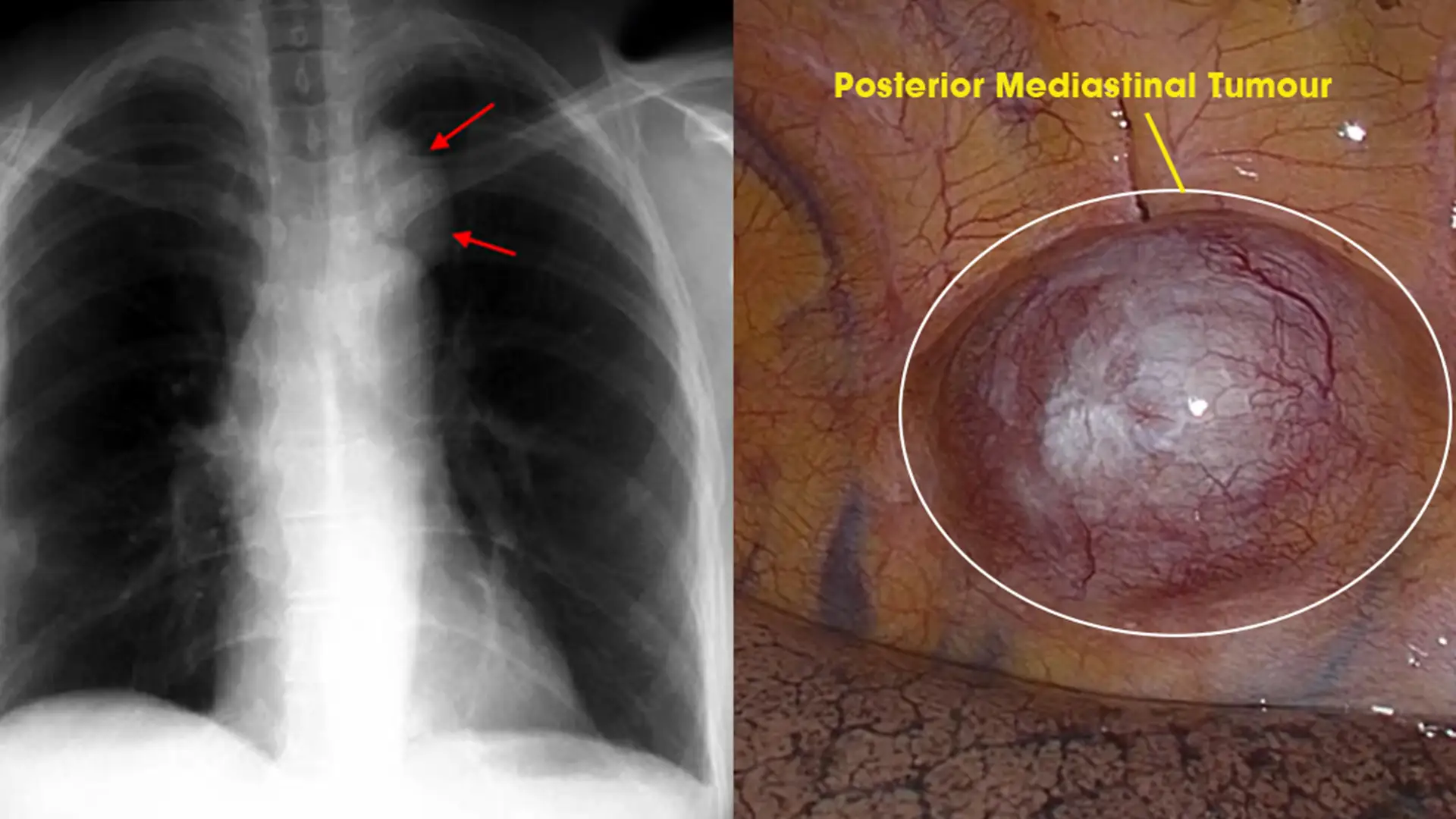
Posterior Mediastinal tumors are benign o...
Copyright @ (Prof.) Dr. Arvind Kumar. All Rights Reserved / Thoracic Surgical Oncologis
License Number: U.P State Medical Council (India) No. 27637


.webp)
 +91-9773635888
 Contact