
MBBS (AIIMS), MS (Surgery, AIIMS), MNAMS, FACS (USA), FICS (USA), FUICC
Mon, 21 Sep 2026
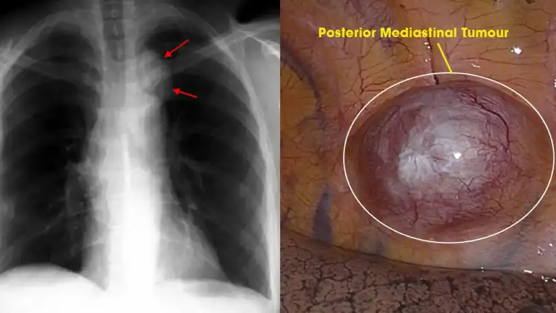
The mediastinum is the central compartment of the thoracic cavity, located between the pleural sacs of the lungs, and houses some of the body’s most vital structures — the heart, esophagus, trachea, and major blood vessels. A tumor in the posterior region of this compartment often presents unique clinical challenges that call for specialized thoracic expertise.
Under the care of Dr. Arvind Kumar, patients receive comprehensive evaluation, accurate diagnosis, minimally invasive treatment options, and personalized post-operative care.
Posterior mediastinal tumors are growths that arise in the back of the chest cavity. Unlike anterior tumors, which are often associated with the thymus gland, tumors in the posterior mediastinum are predominantly neurogenic in origin — they typically develop from the nerves that run alongside the spinal column or from the autonomic nervous system.
Most of these tumors are benign, but their location makes surgery technically challenging: as they grow, they can compress the spinal cord, the esophagus, or the lungs, causing pain or neurological deficits. Approximately 20–30% of mediastinal tumors in adults arise in the posterior mediastinum, and while most are asymptomatic in the early stages, early medical intervention is key to preventing complications.
The first step in posterior mediastinal tumor treatment is identifying the type involved. A few major types are worth knowing:
These are the most frequent type, including Schwannomas, neurofibromas, and ganglioneuromas. While often slow-growing, they can follow the nerve roots through the intervertebral foramina, occasionally creating a “dumbbell” shape that compresses the spinal cord.
Duplication cysts or esophageal diverticula can sometimes mimic a solid tumor in the posterior mediastinum — more on the esophagus and how it is evaluated is covered separately.
Enlarged lymph nodes due to infections, tuberculosis, sarcoidosis, or lymphoma may also present as posterior mediastinal masses.
In rare cases of chronic anemia, the body may attempt to produce blood cells in the posterior mediastinum, forming masses that resemble tumors.
Dr. Arvind Kumar and his team prioritize a comprehensive analysis of symptoms and thorough testing to pinpoint the exact type and location of a posterior mediastinal tumor, so a personalized treatment plan can be built accordingly.
Symptoms vary depending on the size of the mass and its proximity to surrounding structures. In many cases, these tumors are discovered incidentally during a routine chest X-ray or CT scan for unrelated issues; when symptoms do occur, they are often the result of local compression.
These symptoms are often mild and nonspecific, so many patients initially overlook them. As the tumor expands, it may push against the lungs or the esophagus, causing a persistent, dull cough, shortness of breath, or difficulty swallowing if the esophagus is compressed — which can, in turn, contribute to unexplained weight loss and nutritional deficiencies.
Because posterior mediastinal tumors are often neurogenic, they can also cause back pain or intercostal neuralgia. If a tumor begins to invade the spinal canal, patients may experience:
These symptoms require immediate assessment and medical intervention — Dr. Arvind Kumar emphasizes prompt consultation if they arise.
If you are experiencing persistent chest discomfort, chronic cough, unexplained breathing issues, swallowing difficulties, or abnormal imaging findings, consulting an experienced thoracic specialist is important when:
The earlier the condition is diagnosed, the better the treatment outcomes tend to be — schedule a consultation with Dr. Arvind Kumar as soon as alarming symptoms appear.
A proper diagnosis is key to assessing the tumor’s size, shape, and location, which in turn allows for the right surgical approach for complete resection. The following diagnostic tools are commonly used:
This is the most commonly used diagnostic imaging tool, providing detailed cross-sectional views that show the size, density, and exact location of the tumor.
For posterior tumors, an MRI is often preferred over a CT scan because it offers better resolution of soft tissues and can clearly show if a tumor has entered the spinal canal or is pressing against the spinal cord.
Although posterior mediastinal tumors are, for the most part, benign, there is a chance of malignancy. If malignancy is suspected, a PET-CT helps determine whether the tumor is localized or has spread to other parts of the body.
While imaging provides a roadmap, a tissue diagnosis is sometimes required; for many neurogenic tumors that appear benign on imaging, surgeons may proceed directly to excision instead. If a biopsy is necessary, Endobronchial Ultrasound (EBUS) or CT-guided fine-needle aspiration (FNA) may be used. Doctors may also recommend pulmonary function tests (PFTs) to assess lung function and confirm the patient is a fit candidate for surgery.
Dr. Arvind Kumar puts a thorough diagnostic workup at the forefront of his treatment approach before the next steps in the treatment plan are decided.
The primary treatment for most posterior mediastinal tumors is surgical resection, aiming for complete removal of the mass while preserving the surrounding nerves and organs. Thoracotomy was once the standard surgical approach; advances in minimally invasive thoracic surgery now allow many posterior mediastinal tumors to be removed using VATS or robotic surgery, avoiding the need for a large thoracotomy in selected patients.
Video-Assisted Thoracic Surgery (VATS) has changed the treatment of mediastinal tumors. Through three small incisions, a surgeon inserts a camera and specialized instruments to dissect the tumor. This approach reduces blood loss, lowers the risk of infection, and allows patients to return to daily life faster than traditional open surgery.
Using systems like the Da Vinci robot, surgeons get a 3D, high-definition view of the posterior mediastinum with up to 10x magnification. The robotic arms offer a greater range of motion than the human hand, which is valuable when working in the tight, narrow spaces of the posterior chest.
For neurogenic tumors located near the spine, robotic surgery allows for meticulous dissection of the tumor away from the nerves, reducing the risk of neurological damage. Dr. Arvind Kumar has experience performing these robotic techniques for posterior mediastinal tumors that require precise surgical dissection.
When a posterior mediastinal tumor is malignant, the treatment approach differs from that of a benign tumor. Here is a breakdown:
| Treatment Option | Purpose | When It Is Recommended |
|---|---|---|
| Surgery | Complete removal of the tumor and symptom relief | Most benign and operable malignant tumors |
| Video-Assisted Thoracoscopic Surgery (VATS) | Minimally invasive tumor removal with faster recovery | Small to medium-sized tumors without extensive spread |
| Robotic Thoracic Surgery | High-precision minimally invasive surgery | Complex tumors in difficult-to-access areas |
| Open Thoracic Surgery (Thoracotomy) | Safe removal of large or invasive tumors | Tumors involving vital organs, nerves, or blood vessels |
| Chemotherapy | Destroy cancer cells and control tumor growth | Lymphoma, neuroblastoma, or advanced malignant tumors |
| Radiation Therapy | Shrink tumors and reduce recurrence risk | Inoperable tumors or post-surgery cancer management |
| Targeted Therapy / Immunotherapy | Attack specific cancer pathways | Selected advanced cancers based on molecular testing |
| Follow-Up Monitoring | Detect recurrence and assess recovery | Essential after all major treatments |
Dr. Arvind Kumar and his team first diagnose the condition and pinpoint the exact location of the tumor, then determine the most appropriate treatment or surgical plan.
Posterior mediastinal tumors are complex, but with modern medical advancements, they are highly treatable. Whether the tumor is a benign Schwannoma or a more complex malignant growth, the focus remains on complete resection and preserving quality of life. Dr. Arvind Kumar and his team provide minimally invasive care for complex chest conditions, including posterior mediastinal tumor treatment.
For more details about posterior mediastinal tumor treatment and to schedule a consultation, please get in touch with our team directly.

Dr. Arvind Kumar is a highly accomplished Chest surgeon with experience of over 20,000 major chest surgeries, including open surgery, VATS(Video assisted Thoracic Surgery), Robotic Chest Surgery, and Lung Transplantation, reflecting decades of clinical excellence and leadership in chest surgery.
Copyright @ (Prof.) Dr. Arvind Kumar. All Rights Reserved / Thoracic Surgical Oncologist
License Number: U.P State Medical Council (India) No. 27637


 Contact
 +91-9773635888