Lung Cancer Diagnosis
Fri, 15 Dec 2023
Lung Cancer Diagnosis and Screening Tests
We aim to provide innovative lung cancer treatments with latest technologies-all under one roof. We use state-of-the-art diagnostic tools, including advanced imaging and laboratory tests, to evaluate lung cancer. This diagnostic evaluation takes about three to five days. Then together, we chalk out a comprehensive lung cancer treatment plan that works for you together with integrative oncology services to help reduce side effects and keep you strong in physically, mentally and spiritually.
Lung Cancer Diagnosis
Doctors use a wide range of diagnostic procedures and tests to diagnose lung cancer. These include the following:
- The history and physical examination may reveal the presence of symptoms or signs that are suspicious for lung cancer.
- Chest X Ray is the most common first diagnostic step when any new symptoms of lung cancer are present.
- CT (computerized tomography, computerized axial tomography, or CAT) scans may be performed on the chest, abdomen, and/or brain to examine for both metastatic and lung tumors. A CT scan of the chest may be ordered when X-rays do not show an abnormality or do not yield sufficient information about the extent or location of a tumor.
One advantage of CT scans is that they are more sensitive than standard chest X-rays in the detection of lung nodules, that is, they will demonstrate more nodules. Sometimes intravenous contrast material is given prior to the scan to help delineate the organs and their positions. A CT scan exposes the patient to a minimal amount of radiation.
- Low-dose helical CT scan (or spiral CT scan) is sometimes used in screening for lung cancers. This procedure requires a special type of CT scanner and has been shown to be an effective tool for the identification of small lung cancers in smokers and former smokers.
- Magnetic resonance imaging (MRI) scans may be appropriate when precise detail about a tumors location is required. People with heart pacemakers, metal implants, artificial heart valves, and other surgically implanted structures cannot be scanned with an MRI because of the risk that the magnet may move the metal parts of these structures.
- Positron emission tomography (PET) scanning is a specialized imaging technique that uses short-lived radioactive drugs to produce three-dimensional coloured images of those substances in the tissues within the body. While CT scans and MRI scans look at anatomical structures, PET scans measure the function of tissues. PET scans can determine whether a tumor tissue is actively growing and can aid in determining the type of cells within a particular tumor.
- Bone scans are used to create images of bones on a computer screen or on film. Doctors may order a bone scan to determine whether a lung cancer has spread (metastasized) to the bones.
- Sputum cytology: The diagnosis of lung cancer always requires confirmation of malignant cells by a pathologist, even when symptoms and X-ray studies are suspicious for lung cancer. The simplest method to establish the diagnosis is the examination of sputum under a microscope. This is the most risk-free and inexpensive tissue diagnostic procedure, but its value is limited since tumor cells will not always be present in sputum even if a cancer is present.
- Bronchoscopy: Examination of the airways (wind pipe and its divisions) by bronchoscopy (visualizing the airways through a thin, fiberoptic probe inserted through the nose or mouth) may reveal areas of tumor that can be sampled (biopsied) for diagnosis by a pathologist.
- Needle biopsy: Fine needle aspiration (FNA) through the skin, most commonly performed with radiological imaging for guidance, may be useful in retrieving cells for diagnosis from tumor nodules in the lungs.
Needle biopsies are particularly useful when the lung tumor is peripherally located in the lung and not accessible to sampling by bronchoscopy. A small risk (3%-5%) of an air leak from the lungs (called a pneumothorax, which can easily be treated) accompanies the procedure.
- Thoracocentesis: Sometimes lung cancers involve the lining tissue of the lungs (pleura) and lead to an accumulation of fluid in the space between the lungs and chest wall (called a pleural effusion). Aspiration of a sample of this fluid with a thin needle (thoracentesis) may reveal the cancer cells and establish the diagnosis. As with the needle biopsy, a small risk of a pneumothorax is associated with this procedure.
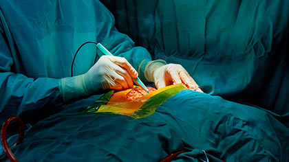
- Major surgical procedures: If none of the aforementioned methods yields a diagnosis, surgical methods must be employed to obtain tumor tissue for diagnosis. These can include.
- Mediastinoscopy (examining the chest cavity between the lungs through a surgically inserted probe with biopsy of tumor masses or lymph nodes that may contain metastases)
- Thoracoscopic Biopsy
- Blood tests: While routine blood tests alone cannot diagnose lung cancer, they may reveal biochemical or metabolic abnormalities in the body that accompany cancer.


.webp)
 +91-9773635888
 Contact